
Diagnosis of cholesteatoma by the b1000 value DWI MRI according to the signal intensity Diagnosis of Cholesteatoma by DWI
Article Sidebar
Main Article Content
Abstract
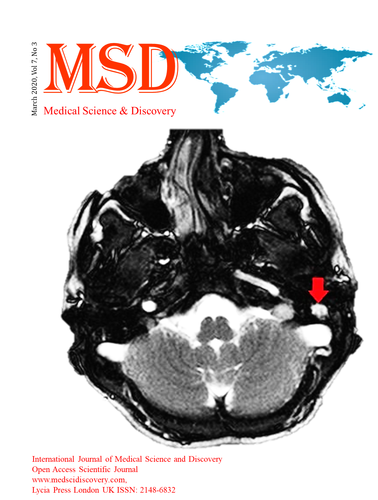
Objective: The cholesteatoma (CL) can be evaluated visually or numerically on an apparent diffusion coefficient (ADC) map, which obtained from at least two different b-valued diffusion-weighted imaging (DWI). In this study, we aimed to evaluate the signal intensity (SI) of the lesion both visually and numerically only on the DWI image without ADC. In case of positive results a second ‘b’ value is not required, so this method could be shorten the duration of the MRI examination.
Material and Methods: Between January 2017 and May 2018, we included patients with chronic otitis media (COM) with a clinical suspicion of primary CL who underwent DWI. Two radiologists and one ear, nose, throat specialist evaluated the radiological images and the pathology results.
Results: The mean SI measurement was significantly higher in the CL group by both observers (observer LR; CL: 107.94 ± 53.36, COM: 37.34 ± 14.70, observer FC; CL: 108.56 ± 50.00, COM: 37.06 ± 15.44; p<0001). ROC analysis showed that a mean SI value of 48.6 was the cut-off value in predicting the diagnosis of CL. The mean SI was significantly higher in the CL group (p<0.001).
Conclusion: We demonstrated a significant difference between CL and COM concerning the diagnosis by visual and numerical signal evaluation only via b1000 valuable images. In false-positive cases, ADC is still confirmatory for high diagnostic accuracy.
Downloads
Article Details
Accepted 2020-03-13
Published 2020-03-22
References
Schwartz KM, Lane JI, Bolster BD Jr, Neff BA. The utility of diffusion-weighted imaging for cholesteatoma evaluation. AJNR Am J Neuroradiol. 2011;32(3):430-436.
Khemani S, Lingam RK, Kalan A, Singh A.The value of non-echo planar HASTE diffusion-weighted MR imaging in the detection, and prediction of the extent of postoperative cholesteatoma. Clin Otolaryngol. 2011;36(4):306-312.
Özgen B, Bulut E, Dolgun A, Bajin MD, Sennaroğlu L. Accuracy of turbo spin-echo diffusion-weighted imaging signal intensity measurements for the diagnosis of cholesteatoma. Diagn Interv Radiol. 2017;23(4):300-306.
Li PM, Linos E, Gurgel RK, Fischbein NJ, Blevins NH. Evaluating the utility of non-echo-planar diffusion-weighted imaging in the preoperative evaluation of cholesteatoma: a meta-analysis. Laryngoscope. 2013;123(5):1247-1250.
Baráth K, Huber AM, Stämpfli P, Varga Z, Kollias S. Neuroradiology of cholesteatomas. AJNR Am J Neuroradiol. 2011;32(2):221-229.
Clarke SE, Mistry D, AlThubaiti T, Khan MN, Morris D, Bance M. Diffusion-Weighted Magnetic Resonance Imaging of Cholesteatoma Using PROPELLER at 1.5T: A Single-Centre Retrospective Study. Can Assoc Radiol J. 2017;68(2):116-121.
Dremmen MH, Hofman PA, Hof JR, Stokroos RJ, Postma AA. The diagnostic accuracy of non-echo-planar diffusion-weighted imaging in the detection of residual and residual cholesteatoma of the temporal bone. AJNR Am J Neuroradiol. 2012;33(3):439-444.
De Foer B, Vercruysse JP, Bernaerts A, et al. Officers detection of postoperative residual cholesteatoma with non-echo-planar diffusion-weighted magnetic resonance imaging. Otol Neurotol. 2008;29(4):513-517.
Keeler JA, Kaylie DM. Cholesteatoma: Is a second stage necessary? Laryngoscope 2016;126(7):1499–1500.
Lingam RK, Khatri P, Hughes J, Singh A. Apparent diffusion coefficients for detection of postoperative middle ear cholesteatoma on non-echo-planar diffusion-weighted images. Radiology 2013;269(2):504–510.
Thiriat S, Riehm S, Kremer S, Martin E, Veillon F. Apparent diffusion coefficient values of middle ear cholesteatoma differ from abscess and cholesteatoma admixed infection. AJNR Am J Neuroradiol 2009;30(1):1123–1126.
Mas-Estelles F, Mateos-Fernandez M, Carrascosa-Bisquert B, et al. Contemporary non-echo-planar diffusion-weighted imaging of middle ear cholesteatomas. Radiographic 2012; 32(3):1197–1213.
Jindal M, Riskalla A, Jiang D, Connor S, O’Connor. A systematic review of diffusion-weighted magnetic resonance imaging in the assessment of postoperative cholesteatoma. Otol Neurotol 2011;32(5):1243–1249.
van Egmond SL, Stegeman I, Grolman W, Aarts. A systematic review of non-echo planar diffusion-weighted magnetic resonance imaging for detection of primary and postoperative cholesteatoma. Otolaryngol Head Neck Surg 2016;15(4):233–240.
Kasbekar AV, Scoffings DJ, Kenway B, et al. Non echo planar, diffusion-weighted magnetic resonance imaging compared with echo planar imaging for the detection of middle ear cholesteatoma. J Laryngol Otol 2011;12(5):376–380.
Akkari M, Gabrillargues J, Saroul N, et al. Contribution of magnetic resonance imaging to the diagnosis of middle ear cholesteatoma: analysis of a series of 97 cases. Eur Ann Otorhinolaryngol Head Neck Dis 2014;13(1):153–158.
Cavaliere M, Di Lullo AM, Caruso A, et al. Diffusion-weighted intensity magnetic resonance in the preoperative diagnosis of cholesteatoma. ORL J Otorhinolaryngol Relat Spec 2014;7(6):212–221.
Dhepnorrarat RC, Wood B, Rajan GP. Postoperative non-echo-planar diffusion-weighted magnetic resonance imaging changes after cholesteatoma surgery: implications for cholesteatoma screening. Otol Neurotol 2009;(30):54–58.
Kosling S, Bootz F. CT and MR imaging after middle ear surgery. Eur J Radiol 2001; 40(2):113–118.
Dubrulle F, Souillard R, Chechin D, Vaneecloo FM, Desaulty A, Vincent C. Diffusion-weighted MR imaging sequence in the detection of postoperative residual cholesteatoma. Radiology 2006;23(8):604–610.
Nash R, Wong PY, Kalan A, Lingam RK, Singh A. Comparing diffusion weighted MRI in the detection of post-operative middle ear cholesteatoma in children and adults. Int J Pediatr Otorhinolaryngol 2015;79(2): 2281–2285.
Suzuki H, Sone M, Yoshida T, et al. Numerical assessment of cholesteatoma by signal intensity on non-EP-DWI and ADC maps. Otol Neurotol 2014;3(5):1007–1010
Garrido L, Cenjor C, Montoya J, Alonso A, Granell J, Gutiérrez-Fonseca R. Diagnostic capacity of non-echo planar diffusion-weighted MRI in the detection of primary and residual cholesteatoma. Acta Otorrinolaringol Esp. 2015 Jul-Aug;66(4):199-204.