
A single center study of oral mucosal lesions in a Turkish population during 12 years period
Article Sidebar
Main Article Content
Abstract
Objective: The prevalence of oral mucosal lesions, together with information on the risk habits associated with oral health, such as tobacco and alcohol use, can help in planning future oral health studies and screening programs.
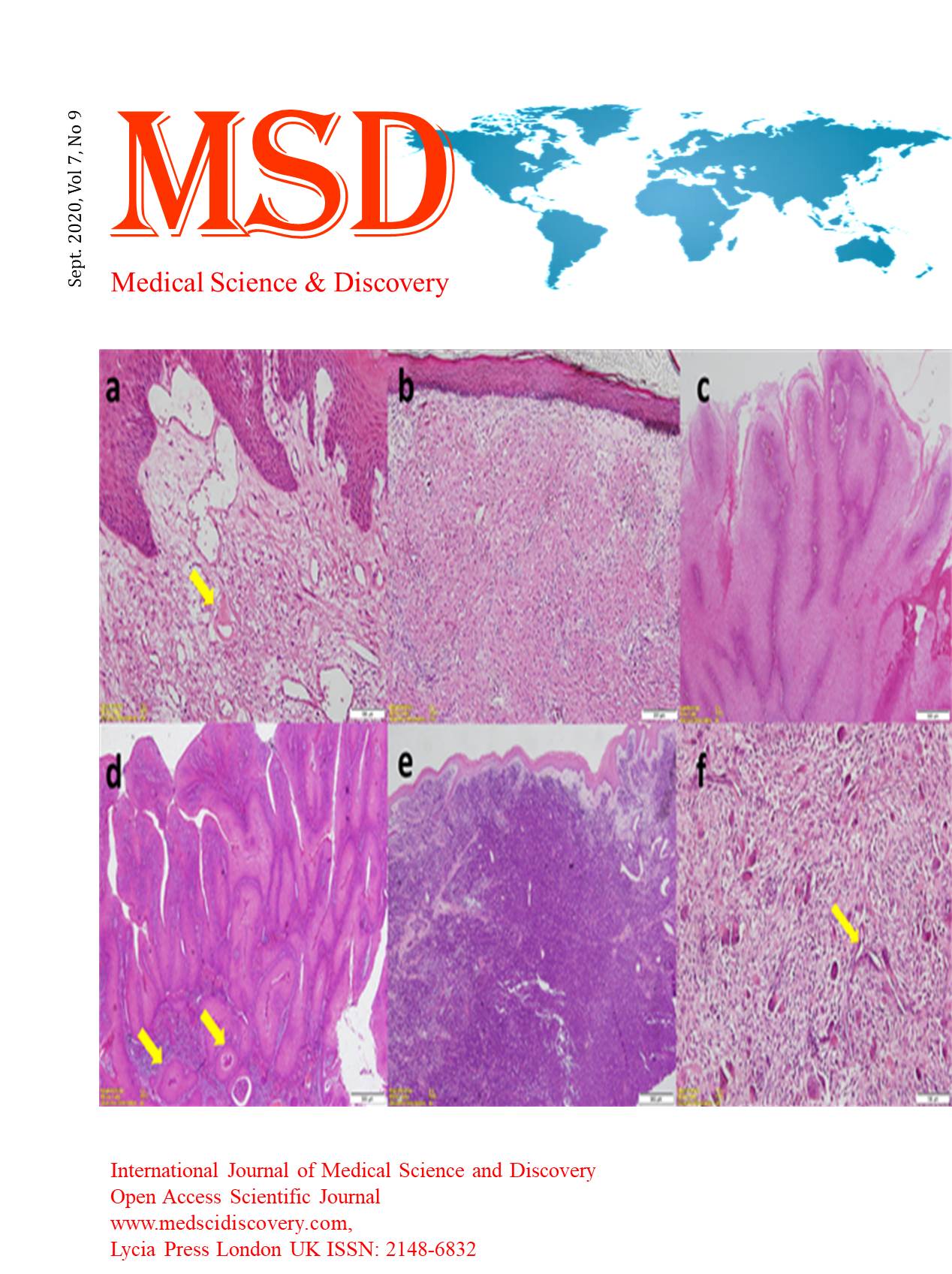
Material and Methods: This study presents the findings of 805 oral mucosal biopsies from patients, received over twelve years period. The cases represent 0.6 per-cent of the total number of reports examined (130.680). The data were revised and compiled for diagnosis site, age, and sex. The patients were divided into nine age groups according to decades. The classification was modified and divided into eleven main groups
Results: Connective tissue lesions formed the largest group of diagnoses (24.4per cent) followed by white lesions (17.8 %per cent), verrucal-papillary lesions (15.4%per cent), red-blue lesions (14%per cent), ulcerous lesions (12.2%per cent), periodontal diseases (10%per cent), lymphoid tissue lesions (1.3%per cent), other tumors (2%per cent), pigmented lesions (0.6%per cent) only 1 metabolic disease (cherubism) (0.1per cent). Approximately 60 %per cent of the biopsies were from the second group patients with an almost equal distribution among sexes. The predominant site of the biopsies was gingiva (28%per cent) followed by lips (19.2%per cent).
Conclusion: The majority of the lesions were in the category of reactive and inflammatory lesions with most occurring in the thirty age group (31-40 age) that represents permanent dentition. These results suggest that the difficulties in maintaining oral hygiene or the presence of trauma may be the primary factor in mucosal lesions occurring in the permanent dentition period.
Downloads
Article Details
Accepted 2020-09-04
Published 2020-09-21
References
Regezi JA, Sciubba J, Jordan J. Oral pathology. Clinical pathologic correlations. 7th ed. St. Louis: Saunders; 2003.
Karcioglu ZA, Someren A. Practical surgical pathology, Massachusetts:Collamore Press, 1985:262.
Köse O, Güven G, Özmen I, Akgün ÖM, Altun C. The oral mucosal lesions in pre-shool and shool age Turkish children. JEADV 2013;27:e136-37.
Majorana A, Bardellini E, Flocchini P, Amadori F, Conti G. Oral mucosal lesions in children from 0 to 12 years old: ten years' experience. Oral Surg Oral Med Oral Pathol Oral Radiol Endod 2010;110:e13-18.
Silva LVO, Arruda JAA, Martelli SJ, Kato CNAO, Nunes LFM, Vasconselos ACU et al. A multicenter study of biopsied oral and maxillofacial lesions in a Brazilian pediatric population. Braz Oral Res. 2018; 32.
Lima GS, Fontes ST, Araújo LMA, Etges A, Tarquinio SBC, Gomes APN. A survey of oral and maxillofacial biopsies in children. A single-center retrospective study of 20 years in Pelotas-Brazil. J Appl Oral Sci 2008;16:397-402.
Parlak AH, Koybasi S, Yavuz T, Yesildal N, Anul H, Aydogan I, et al. Prevalence of oral lesions in 13-to 16-year-old students in Düzce, Turkey. Oral Diseases 2006;12:553-58.
Gültekin SE, Tokman B, Türkseven, MR. A review of paediatric oral biopsies in Turkey. Int Dental J 2003;53:26-32.
Shulman JD. Prevalence of oral mucosal lesions in children and youths in the USA. Int J Paediatric Dentistry 2005;15:89-97.
Almoznino G, Zadik Y, Vered M, Becker T, Yahalom R, Derazne E, et al. Oral and maxillofacial pathologies in young- and middle-aged adults. Oral Diseases 2015;21:493–500.
Carvalho M de V, Iglesias DPP, do Nascimento GJF, Sobral APV. Epidemiological study of 534 biopsies of oral mucosal lesions in elderly Brazilian patients. Gerodontology 2011;28:111–15.
Espinoza I, Rojas R, Aranda W, Gamonal J. Prevalence of oral mucosal lesions in elderly people in Santiago, Chile. J Oral Pathol Med 2003;32:571-75.
Cueto A, Martinez R, Niklander S, Deichler J, Barraza A. Prevalence of oral mucosal lesions in an elderly population in the city Valparaiso, Chile. Gerodontology 2013;30:201-06.
Souza S, Alves T, Santos J, Oliveira M. Oral lesions in elderly patients in referral centers for oral lesions of Bahia. Int Arch Otorhinolaryngol 2015;19:279–285.
Avcu N, Ozbek M, Kurtoglu D, Kurtoglu E, Kansu O, Kansu H. Oral findings and health status among hospitalized patients with physical disabilities, aged 60 or above. Arch Gerontology Geriatrics 2005;41:69–79.
Al-Maweri SA, Al-Jamaei A, Saini R, Laronde DM, Sharhan A. White oral mucosal lesions among the Yemeni population and their relation to local oral habit. J Invest Clin Dent 2018;1-9.
De Giorgi V, Sestini S, Bruscino N, Janowska A, Grazzini M, Rossari S, et al. Prevalence and distribution of solitary oral pigmented lesions: a prospective study. JEADV 2009;23:1320-23.
Hassona Y, Sawair F, Al-Karadsheh O, Scully C. Prevalence and clinical features of pigmented oral lesions. Int J Dermatol 2016;55:1005-13.
Avcu N, Kanli A. The prevalence of tongue lesions in 5150 Turkish dental outpatients. Oral Diseases 2003;9:188-95.
Kovač-Kavčič M, Skalerič U. The prevalence of oral mucosal lesions in a population in Ljubljana, Slovenia. J Oral Pathol Med 2000;29:331–35.
Cebeci ARI, Gülşahı A, Kamburoğlu K, Orhan BK, Öztaş B. Prevalence and distribution of oral mucosal lesions in an adult Turkish population. Med Oral Path Oral Cir Bucal 2009;14: E272-77.
Robledo-Sierra J, Mattson U, Svedensten T, Jontell M. The morbidity of oral mucosal lesions in an adult Swedish population. Med Oral Pathol Oral Cir Bucal 2013;18: E766-72.
Do LG AJ Spencer AJ, Dost F, Farah CS. Oral mucosal lesions: findings from the Australian National Survey of Adult Oral Health. Australian Dental J 2014; 59: 114–20.
Kilinc A, Saruhan N, Gundogdu B, Yalcin E, Ertas U, Urvasizoglu G. Benign tumors and tumor-like lesions of the oral cavity and jaws: An analysis of 709 cases. Niger J Clin Pract 2017;20:1448-54.
Monteiro LS, Albuquerque R, Paiva A, de la Pena-Moral J, Amaral JB, Lopes CA. A comparative analysis of oral and maxillofacial pathology over a 16-year period, in the north of Portugal. International Dental J 2017;67:38–45.
Kansky AA, Didanovic V, Dovsak T, Brzak BL, Pelivan I, Terlevic D. Epidemiology of oral mucosal lesions in Slovenia. Radiol Oncol 2018;52:263-66.
da Silva KD, Rosa WLO, Sarkis‐Onofre R, et al. Prevalence of oral mucosal lesions in population‐based studies: A systematic review of the methodological aspects. Community Dent Oral Epidemiol. 2019;47:431-40.
Mota-Ramírez A, Silvestre FJ, Simó JM. Oral biopsy in dental practice. Med Oral Pathol Oral Cir Bucal 2007;12:E504-10.