
Cutaneous sporotrichosis with dermoscopic features: A case report
Article Sidebar
Main Article Content
Abstract
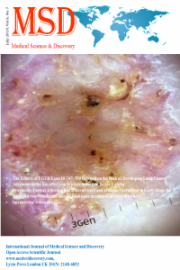
Objective: Sporotrichosis is a fungal skin infection caused by Sporothrix schenckii. We report a 70- year-old female presenting with an erythematous nodule on her right forefinger after a rose thorn prick.
Material and Methods: The nodule was histopathologically diagnosed as sporotrichosis. Dermoscopy of the nodule revealed structureless white areas with a lobular arrangement, prominent scaling, blood spots, and polymorphous vessels including coiled, punctate and looped vessels. Dermoscopy has opened a new horizon in the diagnosis of skin infections in recent years.
Conclusion: The dermoscopic features of sporotrichosis have not yet been reported as far as we are aware. We believe that coexistence of the above features, which could be thought to be nonspecific when seen separately, may be of diagnostic significance and a helpful tool in the diagnosis of cutaneous sporotrichosis.
Downloads
Article Details
References
2. Mahlberg MJ, Patel R, Rosenman K, Cheung W, Wang N, Sanchez M. Fixed cutaneous sporotrichosis. Dermatol Online J. 2009;15:5.
3. Orofino-Costa R, Macedo PM, Rodrigues AM, Bernardes-Engemann AR. Sporotrichosis: an update on epidemiology, etiopathogenesis, laboratory and clinical therapeutics. An Bras Dermatol. 2017;92:606-20.
4. de Lima Barros MB, de Almeida Paes , de Oliveira Schubach A. Sporothrix schenckii and Sporotrichosis. Clin Microbiol Rev. 2011;24:633-54.
5. Zalaudek I, Giacomel J, Cabo H, Di Stefani A, Ferrara G, Hofmann-Wellenhof R. Entodermoscopy: a new tool for diagnosing skin infections and infestations. Dermatology. 2008;216:14-23.
6. Lallas A, Giacomel J, Argenziano G, García-García B, González-Fernández D, Zalaudek I, Dermoscopy in general dermatology: practical tips for the clinician. Br J Dermatol. 2014; 170:514-26.
7. Ayhan E, Ucmak D, Akkurt Z. Vascular structures in dermoscopy. An Bras Dermatol. 2015;90:545-53.
8. Ayhan E, Ucmak D, Baykara SN, Akkurt ZM, Arica M. Clinical and dermoscopic evaluation of cutaneous leishmaniasis. Int J Dermatol. 2015;54:193-201.
9. Ferrari A, Buccini P, Silipo V, De Simone P, Mariani G, Marenda S, Eccrine poroma: a clinical-dermoscopic study of seven cases. Acta Derm Venereol. 2009;89:160-4.