
A Rare Giant Cavernous Hemangioma: A Case Report with the Review of the Literature
Article Sidebar
Main Article Content
Abstract
Objective: Vascular tumors of the breast are extremely rare; among these tumors, hemangiomas are tumors without pathognomonic features. Hemangiomas of the breast are incidentally seen in mastectomy cases. Diagnosis of these tumors using conventional imaging methods is rare. Core needle biopsy and aspiration biopsy cannot help the diagnosis.
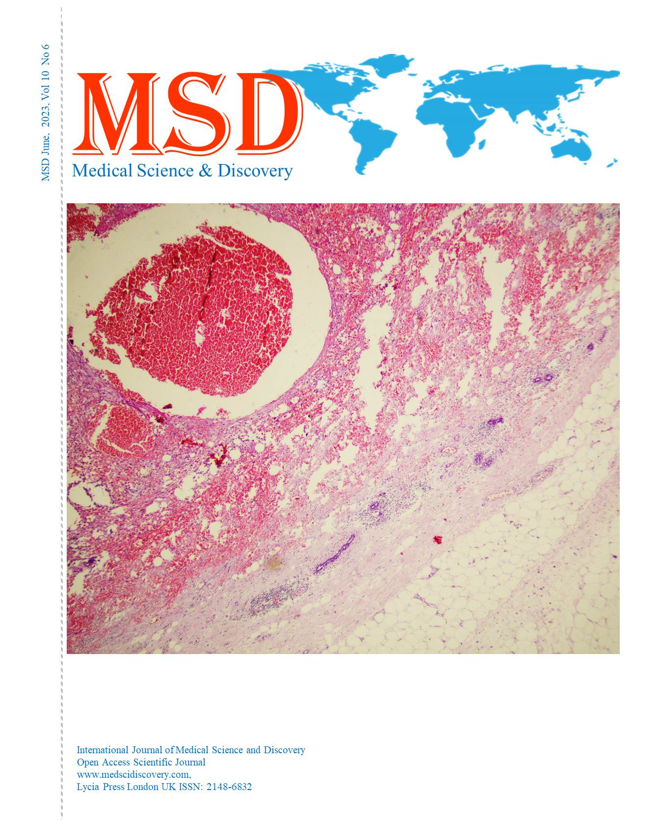
Case: A 69-year-old woman presented to the outpatient clinic with a sudden hematoma on a slow-growing mass in the right breast with a 1-year history. After breast ultrasonography and dynamic contrast-enhanced magnetic resonance imaging supported a 5 × 4 × 2 cm hemangioma, fine needle aspiration biopsy was performed and sent to pathology after excisional biopsy because of the hematinic material. The pathological findings of the mass were positive for breast hemangioma, and no disease recurrence has been observed in the last six months.
Conclusions: Preoperative diagnosis of breast hemangiomas is difficult. Surgical excision is recommended if the vascular lesion is larger than 2 cm, shows atypical features on core needle biopsy, or has discordant radiologic and pathologic findings.
Downloads
Article Details

This work is licensed under a Creative Commons Attribution-NonCommercial 4.0 International License.
Accepted 2023-06-16
Published 2023-06-24
References
Tan PH, Ellis I, Allison K, Brogi E, Fox SB, Lakhani S, et al. The 2019 World Health Organization classification of tumours of the breast. Histopathology. 2020;77(2):181-5.
Ballesio L, Ravazzolo N, Angeletti M, Ambrosio ID, Amabile MI, Pasta V. Diagnostic reflections about a case of breast hemangioma in a woman. Eur J Radiology. 2008;67:5-7.
Salemis NS. Sinusoidal hemangioma of the breast: diagnostic evaluation management and literature review. Gland Surg. 2017;6(1):105-9.
Tilve A, Mallo R, Pérez A, Santiago P. Breast hemangiomas: correlation between imaging and pathologic findings. J Clin Ultrasound. 2012;40:512-7.
Conde DM, de Paula ÉC, Jales RM, de Paula HM, de Sousaet JA. A clinically palpable cavernous hemangioma of the breast in an 80- year old woman. Eur J Obstet Gynecol Reprod Biol. 2013;167:236-7.
Chung SY, Oh KK. Mammographic and sonographic findings of a breast subcutaneous hemangioma. J Ultrasound Med. 2002;21:585-8.
Aslan Ö, Oktay A, Serin G, Yeniay L, Aghamirzayev O. Breast Hemangioma Evaluation with Magnetic Resonance Imaging: A Rare Case Report. Eur J Breast Health. 2022;18(2):190-4.
Jesinger RA, Lattin GE Jr, Ballard EA, Zelasko SM, Glassmanet LM. Vascular abnormalities of the breast: arterial and venous disorders, vascular masses, and mimic lesions with radiologicpathologic correlation. Radiographics. 2011;31:E117-36.
Zhang H, Turner BM, Katerji H, Hicks DG, Wanget X. Vascular lesions of the breast: Essential pathologic features and diagnostic pitfalls. Human Pathology Reports. 2021;26:300570.
Funamizu N, Tabei I, Sekine C, Fuke A, Yabe M, Takeyama H, et al. Breast hemangioma with difficulty in preoperative diagnosis: a case report. World J Surg Oncol. 2014;12:313.
Lee SJ, Mahoney MC. Benign findings in Breast MRI. In: Molleran VM, Mahoney MC. editors. Breast MRI. Philadelphia: Elsevier Saunders. 2014:62.
Adwani A, Bees N, Arnaout A, Lanaspre E. Hemangioma of the breast: clinical, mammographic, and ultrasound features. Breast J. 2006;12:271.
Ameen R, Mandalia U, Marr AA, McKensie P. Breast Hemangioma: MR Appearance with Histopathological Correlation. J Clin Imaging Sci. 2012;2:53.
Kawatra V, Lakshmikantha A, Dhingra KK, Gupta P, Khurana N. A rare coexistence of concurrent breast hemangioma with fibroadenoma: a case report. Cases Journal. 2009;2:7005-7.
Glazebrook KN, Morton MJ, Reynolds C. Vascular tumors of the breast: mammographic, sonographic, and MRI appearances. AJR Am J Roentgenol. 2005;184:331-8.
Mesurolle B, Sygal V, Lalonde L, Lisbona A, Dufresne MP, Gagnon JH, et al. Sonographic and mammographic appearances of breast hemangioma. Am J Roentgenol. 2008;191:W17-W22.
Ginter PS, McIntire PJ, Shin SJ. Vascular tumours of the breast: a comprehensive review with focus on diagnostic challenges encountered in the core biopsy setting. Pathology. 2017;49(2):197-214.
Sebastiano C, Gennaro L, Brogi E, Morris E, Bowser ZL, Antonescu CR, et al. Benign vascular lesions of the breast diagnosed by core needle biopsy do not require excision. Histopathology. 2017;71(5):795-804.
Zhang H, Han M, Varma K, Dabbs DJ. Follow-up outcomes of benign vascular lesions of breast diagnosed on core needle biopsy: a study of 117 cases. Breast J. 2019;25(3):401-7.
Lester SC, Hicks DG. Diagnostic Pathology: Breast, 3rd ed., Elsevier, 2021.